Dermatologists Keep Calling These Light Spots a Fungus. On Our Skin, It Often Isn't.
The look-alike that's more common on skin like ours, why the antifungal never worked, and the one-minute test to ask for by name.
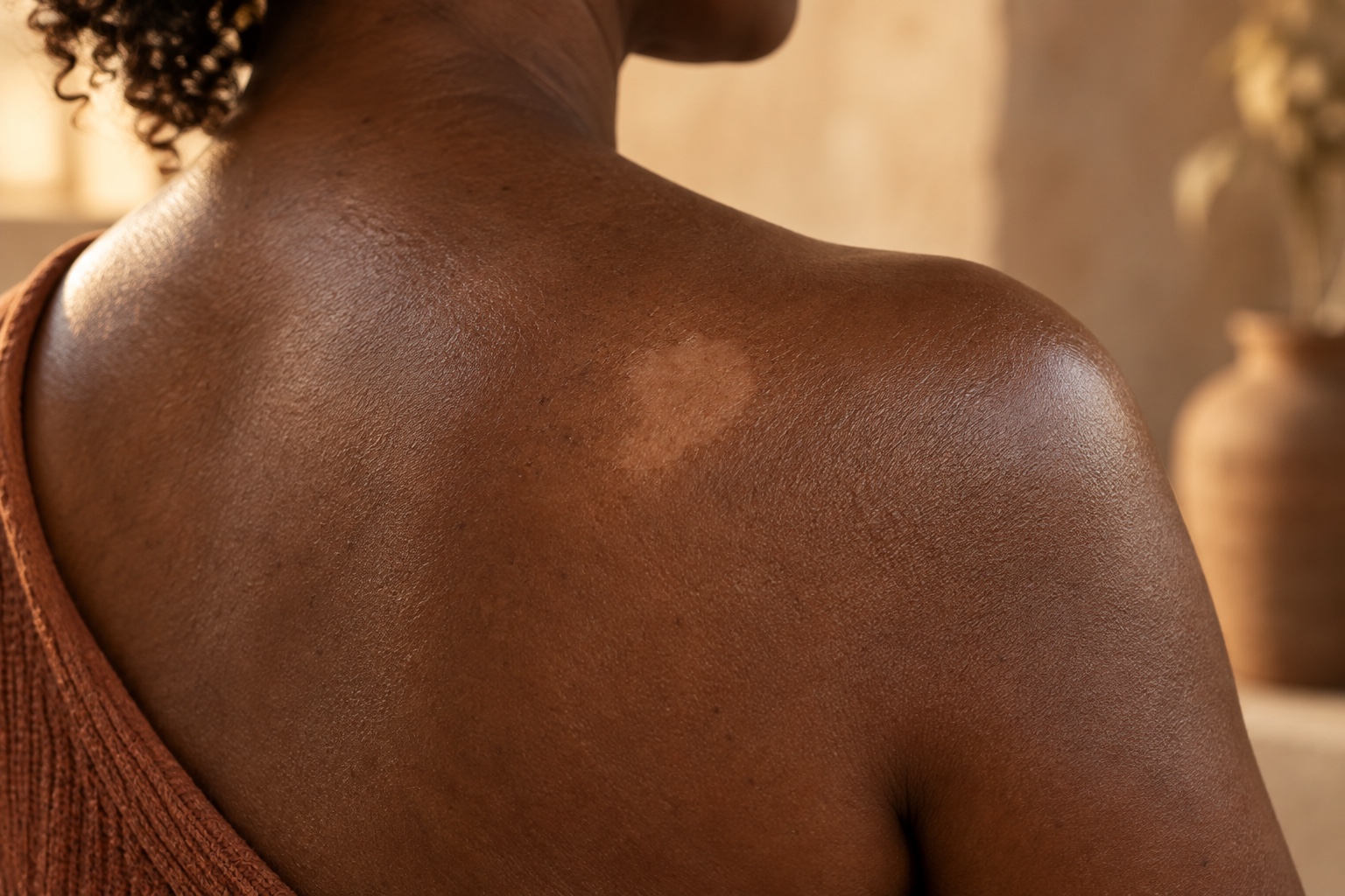
The spots that won't tan
There is a particular kind of light patch that shows up on our skin and quietly ruins a summer. You notice it in July, when everything around it has deepened and these few spots stayed behind — soft-edged, a shade or two lighter than the rest of you, sitting there like the sun skipped them. So you do the responsible thing. You go to a dermatologist. They glance at it, say the word fungus, and send you home with an antifungal.
And then nothing happens. Weeks go by. The cream runs out. The spots don't move.
If that's the story, I want you to know something: you are not imagining it, and you are not doing it wrong. There is a real chance you were handed the correct treatment for the wrong condition — and the reason it happens has a lot to do with the fact that the skin in question is ours.
Two things that look identical and are not the same
The word "fungus" almost always means the dermatologist is thinking of tinea versicolor — an overgrowth of Malassezia, a yeast that lives on everyone's skin and sometimes gets out of hand. It leaves lighter or darker patches, and yes, an antifungal is the right answer for it. On the face of it, a reasonable call.
But there is a second condition that looks so much like tinea versicolor that even dermatologists mix them up constantly, and it is far more common in our skin. It's called progressive macular hypomelanosis, or PMH. The medical literature describes PMH as frequently misdiagnosed and treated inadequately with antifungals or steroids, leaving patients frustrated, and it shows up most in the deeper skin tones, Fitzpatrick four through six — in other words, in us.
Here is the part that matters: PMH is not a fungus. It is driven by ordinary skin bacteria, the same family involved in acne, settling into the hair follicles and quietly turning down the pigment around them. An antifungal cannot touch it. Neither can a steroid. So you can do everything your dermatologist told you, exactly right, for months, and watch it fail — not because skin like ours is stubborn, but because the target was never a fungus in the first place.
Why the summer makes it worse
Both conditions get more obvious in warm months, and the reason is gentler than it looks. The patches themselves usually aren't spreading. It's that the skin around them tans and deepens while the light spots stay put, so the contrast sharpens. Your face didn't get worse. It got more visible. With the fungal version there's an extra wrinkle: the yeast makes a compound that actively suppresses pigment in those spots, so they genuinely won't tan along with everything else.
That's worth holding onto, because "it gets worse every summer" is not evidence that something is wrong with you. It's just contrast doing what contrast does.
How to actually tell them apart, in about a minute
You do not have to guess, and you should not have to spend three months finding out the hard way. There are two quick, cheap, in-office tests that sort this out fast, and it is completely reasonable to ask for them by name.
A Wood's lamp — a handheld ultraviolet light, held over the skin in a dark room. Under it, PMH announces itself: a distinctive coral-red glow coming from the hair follicles, described in the literature as a hallmark of the condition and absent in normal skin. That red glow is a bacterial signal. Fungus does not produce it. Tinea versicolor may show a faint yellowish glow or, often, none at all, which is exactly why the lamp is decisive for PMH and only suggestive for fungus. Vitiligo lights up a bright, chalky white. Pityriasis alba, a mild and mostly-childhood condition, barely glows at all. One lamp, one minute, and the most important question is answered: is that red glow there or not.
A KOH scraping — a painless swipe of the surface, looked at under a microscope. If it's tinea versicolor, the yeast is visible. If it comes back clean in a person of color, that is a flag to think PMH. Standard dermatology guidance is explicit about this: a negative scraping in skin of color should send the doctor's mind toward PMH, and toward treating the bacteria, not the fungus.
And there is a third clue you can check yourself, in the mirror, tonight. PMH lives on the trunk: the chest, the back, the stomach, around the midline. It only rarely reaches the face and neck. So if these light spots are only on the face and nowhere on the torso, PMH becomes a weaker bet, and the more likely culprits are tinea versicolor or old hypopigmentation left behind by acne or irritation that healed lighter. If there are matching spots on the chest or back, PMH climbs the list. That single observation tells your dermatologist more than another appointment of squinting will.
What to do while you sort it out
Some of this is safe to start no matter which condition it turns out to be.
Sunscreen, daily, is the highest-value thing on this list. Not for vanity — the light patches have less pigment, which means less built-in protection, so they burn more easily. And keeping the surrounding skin from tanning darker is what flattens the summer contrast that made the spots loud in the first place. Broad-spectrum, every day, on the face and any affected area.
Be gentle. Skip the scrubbing, the hot water, the harsh actives directly on the patches. Irritation on our skin has a way of leaving its own marks, and the last thing a pigment problem needs is a second pigment problem layered on top.
If the antifungal already failed, benzoyl peroxide is the reasonable next thing to try, and this is not a folk remedy. Benzoyl peroxide plus a clindamycin lotion, and in a dermatologist's hands light therapy, is the established treatment for PMH — precisely because it goes after the bacteria the antifungal ignored. Let me be honest about the odds, because you deserve the real number: in the best comparison we have, topical antibacterial treatment helped roughly a third of patients, while light therapy helped about ninety percent but tended to relapse, and about a quarter of people recovered on their own with no treatment at all. So benzoyl peroxide is a legitimate, cheap, evidence-backed move, not a switch that flips it back overnight. Use it gently, since it dries skin and bleaches fabric, moisturize after, and give it real time.
And brace for slow. However this resolves, repigmentation runs on the order of months, sometimes longer. That is normal. A treatment that hasn't worked in two weeks hasn't failed. It's just early.
The one thing to take with you
If a dermatologist looks at light spots on our skin, says "fungus," and reaches for the prescription pad without ever running a Wood's lamp or a scraping, that is the moment to speak up. Not because they're careless, but because the two conditions that look most alike here demand different treatments, and the one that's more common in us is the one that gets missed. Asking for the lamp and the scraping by name is the difference between another season of the wrong cream and actually treating the thing you have.
Our skin is not harder to diagnose. It has just been studied less, pictured less in the textbooks, and second-guessed more. You knowing the name of the test is part of how that changes.
The short version
Light spots that won't tan, a dermatologist who said fungus, a cream that did nothing: there is a second condition that looks identical, is far more common on our skin, and is not a fungus at all. Progressive macular hypomelanosis. Bacteria in the hair follicle, not yeast on the surface. Antifungals and steroids cannot touch it; benzoyl peroxide, clindamycin, and light therapy can. It lives on the trunk, the chest, back, and stomach, and only rarely on the face, so check your torso. Ask for a Wood's lamp and a KOH scraping by name: the lamp glows coral-red for PMH and stays dark for fungus, and the scraping settles the rest in a minute. Sunscreen on the patches every day protects them and keeps the summer contrast from getting louder. Repigmentation is slow. Months, not weeks.
Sources
- Elmariah SB, Kundu RV. Progressive macular hypomelanosis. J Drugs Dermatol. 2011;10(5):502–6. PubMed PMID 21533296.
- Relyveld GN, Menke HE, Westerhof W. Progressive macular hypomelanosis: an overview. Am J Clin Dermatol. 2007;8(1):13–9. https://doi.org/10.2165/00128071-200708010-00002
- Leonard N, Krueger S, Rashighi M. Successful treatment of progressive macular hypomelanosis. Dermatol Reports. 2020;12(2):8509. https://doi.org/10.4081/dr.2020.8509
- Paquette GM, Sherman S, Akabane A, Harris JE. Adolescent extra-truncal progressive macular hypomelanosis. Pediatr Dermatol. 2023;40(4):702–705. https://doi.org/10.1111/pde.15252
- Hwang SW, et al. Progressive macular hypomelanosis in Korean patients: a clinicopathologic study. Ann Dermatol. 2009;21(3):261–7. https://doi.org/10.5021/ad.2009.21.3.261
- Kim MB, et al. Narrowband UVB treatment of progressive macular hypomelanosis. J Am Acad Dermatol. 2011;66(4):598–605. https://doi.org/10.1016/j.jaad.2011.04.010
- Thng STG, Long VSH, Chuah SY, Tan VWD. Efficacy and relapse rates of different treatment modalities for progressive macular hypomelanosis. Indian J Dermatol Venereol Leprol. 2016;82(6):673–676. https://doi.org/10.4103/0378-6323.182797
- Lin RL, Janniger CK. Pityriasis alba. Cutis. 2005;76(1):21–4. PubMed PMID 16144284.
- Wigger-Alberti W, Elsner P. Fluorescence with Wood's light. Hautarzt. 1997;48(8):523–7. https://doi.org/10.1007/s001050050622
- Ankad BS, Dhananjaya S. Erythrasma mimicking tinea versicolor: the diagnostic utility of ultraviolet-induced fluorescence dermoscopy. Cureus. 2025;17(11):e96970. https://doi.org/10.7759/cureus.96970
Reign in your skin
Doctor Djeli is an educational resource, not medical advice.
Evaluate your products →Doctor Djeli is an educational resource, not medical advice.